James Allison, a 2018 Nobel laureate and chair of the department of immunology at MD Anderson Cancer Center, will be the featured speaker at a live, virtual commencement hosted by the Gerstner Sloan Kettering Graduate School of Biomedical Sciences (GSK). Eight students who successfully defended their dissertations this past year will be awarded PhD degrees during the school’s ninth commencement ceremony on May 27 as part of the 41st annual academic convocation at Memorial Sloan Kettering.
“We are excited and honored that Dr. Allison has agreed to participate in our virtual event to celebrate this impressive group of graduates, whose hard work and innovative biomedical research deserves to be recognized and applauded,” says GSK Dean Michael Overholtzer. “Dr. Allison conducted part of his Nobel prize–winning immunology research at MSK, so we are fortunate to have him return and share his unique experiences and insightful outlook with these scientists, who are beginning their professional careers.”
The 2020 commencement is being planned as a virtual event because of the “New York State on Pause” stay-at-home order put in place as a response to the COVID-19 pandemic. The ceremony will recognize students who have completed their thesis work in laboratories at MSK, including those graduating from both GSK and the Weill Cornell Graduate School of Medical Sciences. It will also honor distinguished scientists and clinicians at MSK and beyond for their research success.
GSK will award doctorates to Ivan Cohen, Christopher Hulton, Mark Jelcic, James Keith, Jr., Nicholas Kuhn, Hannah Wise, Brittany Woods, and Yuchen Xie. Their graduation brings the total number of GSK alumni to 75. Below are summaries of their thesis projects.
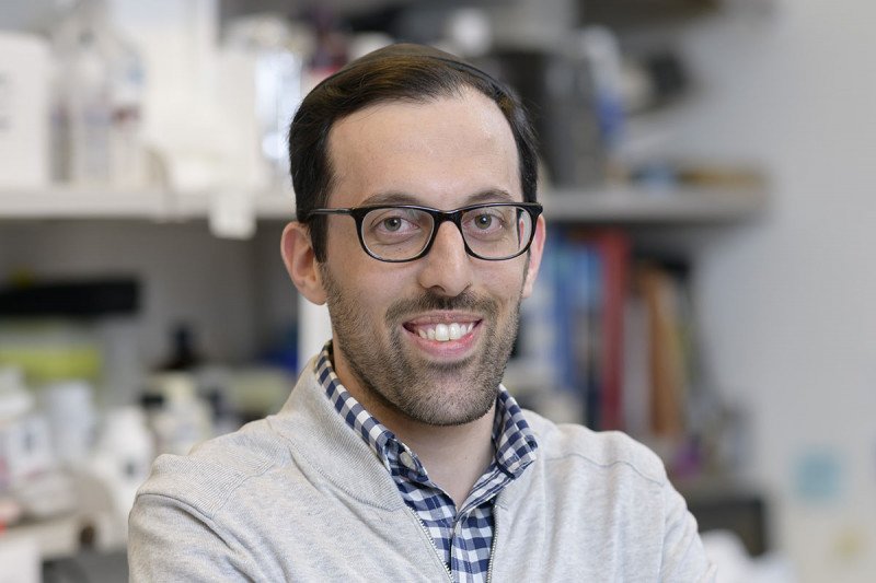
I chose to earn my PhD at GSK because I was confident that it would give me the best opportunity to do meaningful, translational cancer research. I conducted my thesis work in the laboratory of physician-scientist Ronald Blasberg, whose research focuses on the development of noninvasive imaging models in living organisms using radionuclide and optical reporter systems.
My dissertation centers on the role of immune cells in fighting aggressive triple-negative breast cancer. My goal was to gain a better understanding of the relationship between increased lactic acid secretion, known as tumor glycolysis, and immune infiltration of solid tumors, which has long been recognized as critical for long-term survival. In fact, people with immune-infiltrated tumors tend to have much better responses to immune checkpoint blockade therapy.
An abundance of literature shows that lactic acid is highly immunosuppressive in multiple immune cell types. We used cell lines, mouse models, and bioinformatic approaches to study whether glycolysis reduces the infiltration and function of immune cells in breast cancer.
We found that glycolysis, which leads to an increased accumulation of lactic acid outside of the cell, was associated with decreased immune infiltration and function in both animal models of breast cancer and human tumor samples.
I am now a postdoctoral research fellow at the Weizmann Institute of Science in Israel.
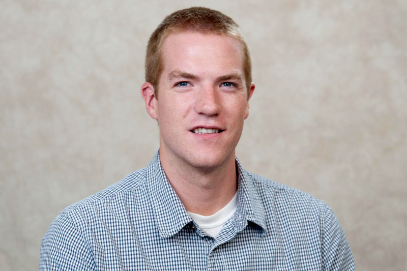
I completed my dissertation project in the lab of physician-scientist Charles Rudin, Chief of the Thoracic Oncology Service at MSK. My research focused on the development of a platform to enable direct genome editing in the tumor models known as patient-derived xenografts (PDXs).
PDXs accurately model many key aspects of human cancer because they are generated from tissue or cells grafted from a patient’s tumor that are then implanted into an immunodeficient mouse. However, PDX use has been limited in cancer research because of technical barriers that do not affect other types of cancer models. As a result, scientists have not been able to perform targeted genome editing of these tumors with existing technologies.
To address this limitation, we developed a new method to perform gene editing of PDXs in mice using CRISPR-Cas9. This system is the first to be developed specifically for use in PDXs and is able to overcome the technical barriers of using this cancer model.
Our research showed how this platform could be used to analyze genes that specific tumor cell lines depend on for survival as well as mechanisms of drug resistance in PDX models of lung cancer. This flexible system has broad application and substantially enhances the utility of PDXs as genetically programmable models of human cancer.
I am now working as a research scientist at C4 Therapeutics.
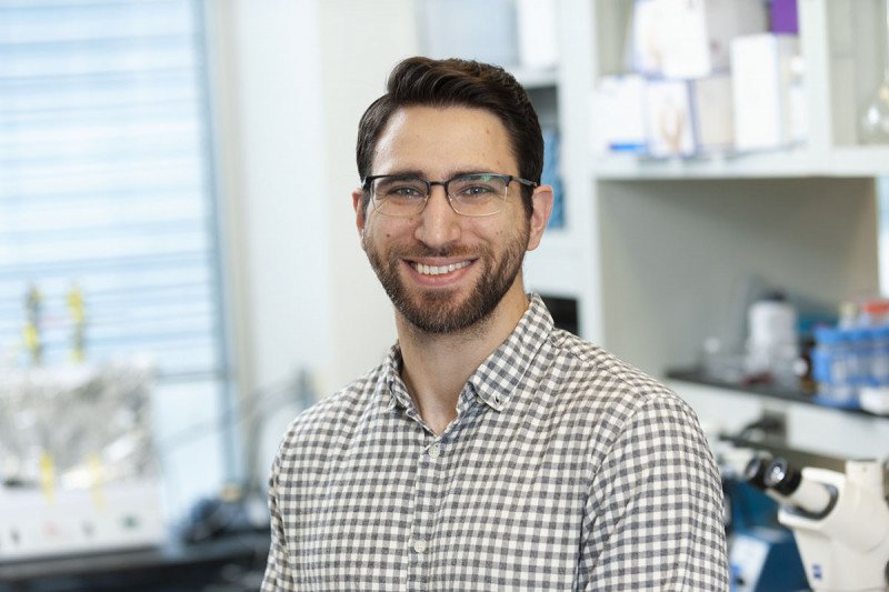
I chose to pursue my doctorate at GSK because of the high-caliber basic science and translational research being conducted at MSK.
I completed my thesis research in the laboratory of cell biologist Philipp Niethammer. His lab uses cutting-edge quantitative imaging techniques to study how tissues detect wounds and initiate healing, inflammation, regeneration, and cancer. The originality and rigor of the science being conducted in Dr. Niethammer’s lab made it an easy decision for me to pursue my graduate training under his guidance.
Adenosine 5’-triphoshate (ATP) is a key signaling molecule in inflammation and wound healing. ATP is an allosteric regulator and ligand, which means that it binds to inactive sites on proteins. However, identifying noncatalytic ATP binding sites on proteins is challenging, and our current inventory of ATP-controlled pathways is likely incomplete. My thesis project focused on using innovative biochemical methods to identify ATP-binding proteins and to further investigate and uncover ATP signaling mechanisms.
I collaborated with the lab of chemical biologist Minkui Luo to develop an original ATP photo-affinity probe that would identify proteins that directly interact with ATP to modulate cell signaling. The probe enabled us to perform comprehensive proteomic screening and find novel ATP-binding proteins on cell membranes. These newly discovered ATP binders suggest that there are still uncharacterized roles for ATP in critical cell processes.
I am now a postdoctoral research fellow at MSK.
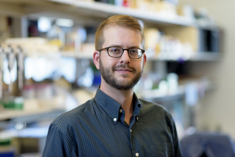
I conducted my dissertation research under the guidance of physician-scientist Eric Pamer. My thesis project examined the impact of intestinal domination by antibiotic-resistant bacteria on the colonic mucosa (the inner lining of the colon and rectum).
Under normal circumstances, the colon is colonized by complex bacterial populations that constitute the microbiota. One function of the microbiota is to provide resistance against intestinal pathogens. Antibiotic treatment of people undergoing complex medical therapies, such as allogeneic hematopoietic cell transplantation, can deplete the gut microbiota, increasing susceptibility to infection by pathogens, such as Clostridioides difficile (C. difficile).
My project focused on whether the residual microbiota after antibiotic treatment influences the severity of colitis (inflammation in the colon) caused by C. difficile. To address this, I colonized antibiotic-treated mice with antibiotic-resistant microorganisms — including vancomycin-resistant Enterococcus faecium, Klebsiella pneumoniae, Escherichia coli, and Proteus mirabilis — and challenged them with C. difficile.
I found that the severity of C. difficile infection and mortality was not significantly influenced by the residual microbiota after antibiotic treatment. These findings indicate that the pathogenetic mechanisms engaged by C. difficile to cause colitis, and the host’s immune defenses leading to resolution, override the relatively minor impact of residual bacterial species on colonic immune defenses.
I completed my thesis work under the guidance of physician-scientist Renier Brentjens, Director of Cellular Therapeutics at MSK. His research focuses on cancer immunology.
My dissertation centers on how the immune system tracks and fights cancer. I specifically sought to discover how T cells, known as the soldiers of the immune system, can be engineered, or manipulated, to strengthen their ability to recognize and eliminate cancer cells. This research topic appealed to me because it required finding creative ways to tweak the immune system in order to ultimately improve patient outcomes.
While the immune system can target and eliminate cancer cells in healthy people, this ability is lost in sick people. Genetic engineering of T cells via chimeric antigen receptors (CARs) is a powerful therapeutic technique that strengthens a person’s own T cells so they can eliminate cancer cells. However, some patients do not respond to this therapy. We used CARs as a jumping-off point to see if, through specific genetic engineering of T cells, we could recruit other types of immune cells to aid in the antitumor response.
We engineered CAR T cells to always express a specific immunological protein: CD40 ligand (CD40L). This strategy rang the metaphorical alarm bells of the immune system when the CAR T cells were engaged in fighting cancer. This led to the recruitment and activation of additional cancer-fighting immune cells that reinforced the CAR T cells so that they did not have to fight alone. Ultimately, this call to arms and the cooperation of immune cells resulted in improved survival in preclinical mouse models.
We concluded that by equipping CAR T cells with CD40L, we can greatly enhance the ability of the immune system to attack cancer cells and thwart their growth. This could potentially increase the number of patients who may benefit from this therapy.
I now work in the laboratory of Matthew Krummel at the University of California in San Francisco.

I chose to pursue a PhD in cancer biology at GSK because I knew I would have a direct impact on patients’ care while working to understand the science behind the disease.
I completed my thesis work in the laboratory of physician-scientist David Solit, Director of the Marie-Josée and Henry R. Kravis Center for Molecular Oncology at MSK. His lab focuses on signaling pathways, which tell a cell when to grow and expand. When these pathways are overactivated, a cell will grow uncontrollably, sometimes leading to cancer.
My work focused on using DNA sequencing to determine the genetic mutations commonly found in bladder cancer. Many of the mutations we observed were in the genes known as epigenetic regulators. Epigenetic regulators control the expression of many genes in a cell, and alterations in these regulators can have widespread effects across the genome.
My research showed that genetic alterations resulting in the loss of one of these regulators, known as KDM6A, can lead to increased tumor development and proliferation in cell lines as well as in mouse models. Additionally, we found that KDM6A mutations were associated with early-stage disease. In contrast, another epigenetic regulator, known as ARID1A, was associated with later-stage disease that had metastasized. This suggests that epigenetic regulators have different functions throughout the course of disease and may be viable targets for therapies in the future.
I now work as a lead clinical data analyst at Flatiron Health, an oncology and technology company in New York.
I conducted my dissertation research in the lab of physician-scientist Ross Levine, Chief of the Molecular Cancer Medicine Service in the Human Oncology and Pathogenesis Program at MSK. My thesis work focused on developing new mouse models of blood cancers caused by mutations in a gene called JAK2. The models revealed fresh insights around the genetic and epigenetic basis of these cancers and how these factors affect disease progression and therapeutic response.
Myeloproliferative neoplasms (MPNs) occur when the bone marrow makes too many red blood cells, platelets, or white blood cells. There are three main kinds of MPNs: polycythemia vera, essential thrombocythemia, and primary myelofibrosis. Despite their clinical differences, all three are driven by genetic mutations in the JAK/STAT signaling pathway.
The most frequent of these events is a V617F mutation in JAK2. Previous studies have revealed that MPNs originate from hematopoietic stem cells, which can develop into different types of blood cells. This suggests that the JAK2 V617F mutation is present in, and likely affects, all hematopoietic lineages, regardless of the MPN subtype.
I generated several mouse models to identify and study factors that influence the role of JAK2 V617F in the development of MPNs. We were particularly interested in studying the role of JAK2 V617F in the megakaryocyte (Mk) lineage. Mks are large bone marrow cells that are responsible for platelet production.
We observed that mutant Mks and platelets contribute to MPN development by promoting increased differentiation of JAK2 wild-type erythroid (red blood cell) lineage cells. This is caused in part by the increased production of interleukin-6 by mutant Mks. Overall, this work highlights a previously underappreciated role for JAK2 V617F in promoting MPN development via cytokine-mediated expansion of the JAK2 wild-type hematopoietic compartment.
I am now a research associate at William Blair & Co., an investment management firm, where I focus on the biotechnology sector.

I conducted my thesis work in the laboratory of physician-scientist Emily Cheng. Her research focuses on the molecular mechanisms of cell death and their implications on tumor development and cancer treatment. Recently, Dr. Cheng has broadened her research to study the epigenetic regulators that are commonly mutated in both kidney and lung cancers.
The gene SETD2 is frequently mutated in a wide variety of human cancers, with the highest mutation rates occurring in clear cell renal carcinoma (ccRCC), the most common and aggressive type of kidney cancer, and lung adenocarcinoma, the most common form of lung cancer. My dissertation centers on how mutations in SETD2 impact the development, spread, and treatment of these diseases.
Previous research has revealed that SETD2 mutations are strongly associated with the metastasis of ccRCC. In addition, SETD2 deficiency has been reported to team up with the genetic mutation KRAS G12D to promote the initiation of lung cancer. However, it remains unclear how SETD2 loss of function supports tumor development.
I developed an SETD2 knockout mouse model and used a patient-derived cell line with SETD2 mutations to investigate the role of SETD2 in the development of lung cancer and the spread of kidney cancer. We found that SETD2 deficiency accelerated the initiation of KRAS G12D–driven lung cancer, increased tumor burden, and significantly reduced mouse survival.
Our results also showed that SETD2 is a strong suppressor of ccRCC tumor metastasis and shed light on the molecular mechanisms that contribute to this problem. We found that in kidney cancer, SETD2 loss of function induced expression of the gene MMP1, which promotes tumor metastasis. Overall, SETD2 loss creates a permissive molecular environment for oncogenes to work together to drive tumor initiation or metastasis.
Furthermore, we uncovered treatment strategies that target and inhibit the mechanisms fueling SETD2-deficient cancers. In both ccRCC and lung cancer cell lines, SETD2 loss sensitized cancer cells to cell death when a family of proteins known as histone chaperones was inactivated through treatment with certain chemical inhibitors. We found that these treatments significantly prolonged the survival of SETD2-deficient mice bearing KRAS G12D–driven lung tumors and suppressed tumor growth in ccRCC mouse models with SETD2 mutations.
I am now a postdoctoral research associate at the New York Genome Center.