In 90% of cancer deaths, what kills is not the primary tumor. Rather, it’s the spreading of cancer to other vital tissues and organs, what’s called metastasis, that proves deadly.
Scientists are eager to better understand this process so they can intervene in it, potentially saving lives.
Cell biologist Joan Massagué has been systematically dissecting metastasis for more than two decades. His research has shown that it is the end result of a long process that can be divided into several steps, each with its own challenges.
The latest study to emerge from his lab identifies the final hurdle that metastatic cancer cells, like microscopic seeds, must overcome to take root and sprout somewhere else.
“We’ve identified a molecule that metastasis-initiating cancer cells need in order to start growing in a new location,” explains Dr. Massagué, who also serves as Director of the Sloan Kettering Institute. “If we deprive the cells of this molecule, they can still infiltrate distant organs, but they fail to grow into a new tumor.”
The findings, published on July 23 in Nature Cell Biology, apply to several different types of cancer as well as different metastatic sites. This suggests a common vulnerability that could be exploited and targeted across cancers.
Cancer Metastasis: A Dangerous Voyage
For cancer cells to metastasize from one part of the body to another, they must successfully traverse a dangerous voyage. From a primary tumor, they must dislodge, enter the blood or lymph circulation, and travel to another part of the body. At this new site, they must squeeze through a blood vessel wall, take up residence in the tissue, and start growing again. Less than 1% of all cancer cells that begin the journey will complete it; most will perish along the way.
Even those that do reach a secondary spot are not guaranteed to grow in the new location. This final step, called colonization, is essentially the last hurdle that metastatic cells must overcome.
“In some ways, it’s the most critical step because what happens next is growth of a new tumor,” says Emrah Er, a postdoctoral fellow in the Massagué lab and the first author on the new paper.
Dr. Er notes that the majority of cancer cells die when they get to the secondary site because they do not know how to cope with the new local conditions. But some do, and it’s those they want to understand better.
More than a Sticky Molecule
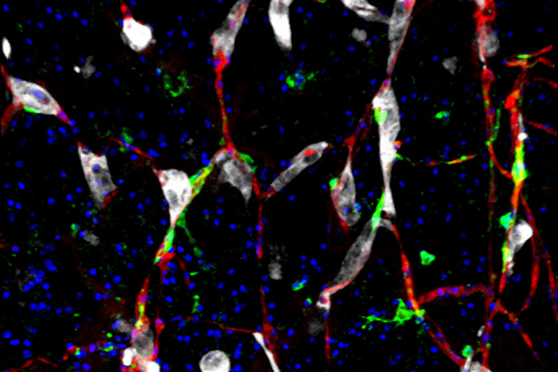
From previous experiments, the researchers knew that cancer cells metastasizing to the brain cling to the surface of blood capillaries there. They had also identified a molecule, called L1CAM, that plays a role in this adhesion.
This molecule is not restricted to brain metastases, however. The researchers also found it in lung, liver, and bone metastases. What’s more, these came from several different primary tumors, including lung, breast, colorectal, and kidney cancers. The presence of L1CAM is associated with recurrence and overall poor survival in these cancers.
That raised the question of just what this molecule is doing to facilitate metastasis in these different cases. To get at an answer to this question, the team used genetic tools to knock down the action of this molecule.
“When we did this, we found that cancer cells were still able to migrate through the blood to new destinations, but once there, they didn’t grow,” explains Dr. Er.

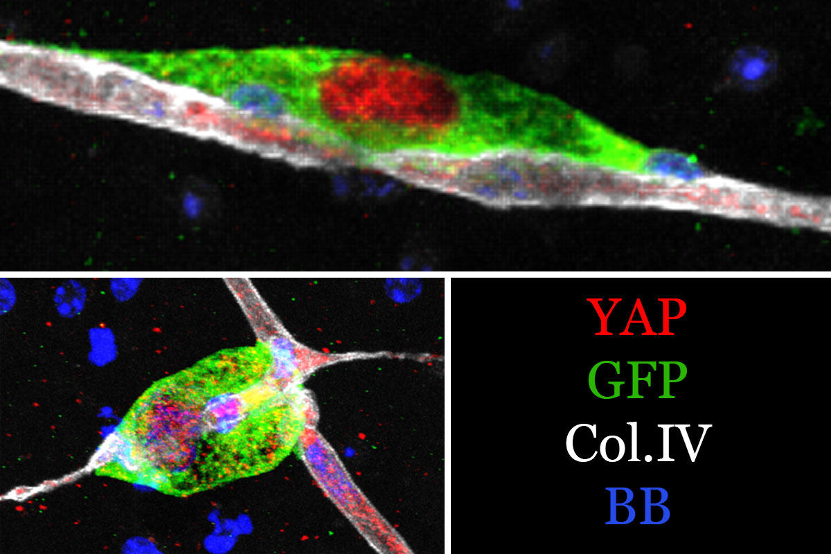
Using advanced imaging technology, the team then watched what cells with working L1CAM did upon arrival to the outer surface of a capillary. The cancer cells spread in an amoeba-like fashion along its length. The behavior reminded the researchers of another cell type that normally coats capillaries: pericytes.
Pericytes wrap around capillaries like insulation around a pipe. By constricting and relaxing, they regulate blood flow through capillaries. When the scientists peered inside the cancer cells with biochemical tools, they discovered that some of the same signals that pericytes use to do their work were active in the cancer cells. Only the cancer cells were using the signals for more nefarious purposes.
These signals, proteins called YAP and MRTF, are known to respond to mechanical changes in tension or force. The researchers believe that the physical act of spreading along a stiff capillary trips these mechanical sensors, providing a necessary cue for growth. And indeed, when the team blocked these sensors, the cancer cells failed to grow.
“Removing YAP and MRTF mimicked the results we obtained when we knocked down L1CAM,” Dr. Er says. “So we know they are the proteins that are critical to this process.”
The results provide a different explanation than the standard one for why metastatic cells cling to capillaries. It’s not just about obtaining oxygen and nutrients, as is conventionally assumed. It’s also about securing anchorage that provides the green light for growth.
The Massagué lab is now determining the origin of these metastasis-initiating cells. They’re also exploring whether it’s possible to intervene in this process by targeting L1CAM with antibodies that can block its function.
An accompanying editorial about the new study appears in the same issue of Nature Cell Biology.


