Quantitative imaging of physical parameters using MRI can be used to improve diagnosis and to assess early treatment response. For example, diffusion MRI can image tissue cellularity and perfusion MRI can map microvasculature, providing valuable biologic information. However, performing quantitative MRI in the body suffers from geometric distortions, sensitivity to motion, and lower signal-to-noise ratio. Our lab is working on the following projects to develop new MRI technology that can enable robust quantitative MRI in the body:
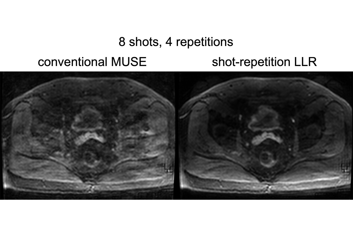
- High-resolution distortion-reduced diffusion MRI using multi-shot acquisition and shot-repetition reconstruction: k-space acquisition is segmented into different shots and repetitions to shorten the readout and thus reduce sensitivity to magnetic field inhomogeneities, while the reconstruction algorithm exploits multidimensional correlations to reduce the amount of data and enable diffusion MRI with higher resolution and reduced distortions without a significant scan time increase. Figure 5 shows preliminary results using a k-space acquisition segmented into 4 shots and 4 repetitions, and a reconstruction algorithm that exploits locally-low-rank along the combined shot-repetition dimension.
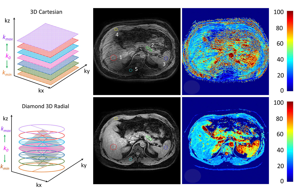
- Fast motion-resistant T1-rho imaging using radial acquisition and fingerprinting-based quantification: radial sampling will enable to image during free-breathing and fingerprinting-based quantification will reduce the amount of data and improve the quantification of T1-rho values for fast motion-resistant T1-rho of abdominal tumors. Figure 6 shows preliminary results of using a fast radial acquisition named “diamond sampling” to perform free-breathing T1-rho imaging in the liver.
Fast motion-resistant T1-rho imaging using radial acquisition and fingerprinting-based quantification: radial sampling will enable to image during free-breathing and fingerprinting-based quantification will reduce the amount of data and improve the quantification of T1-rho values for fast motion-resistant T1-rho of abdominal tumors. Figure 6 shows preliminary results of using a fast radial acquisition named “diamond sampling” to perform free-breathing T1-rho imaging in the liver.