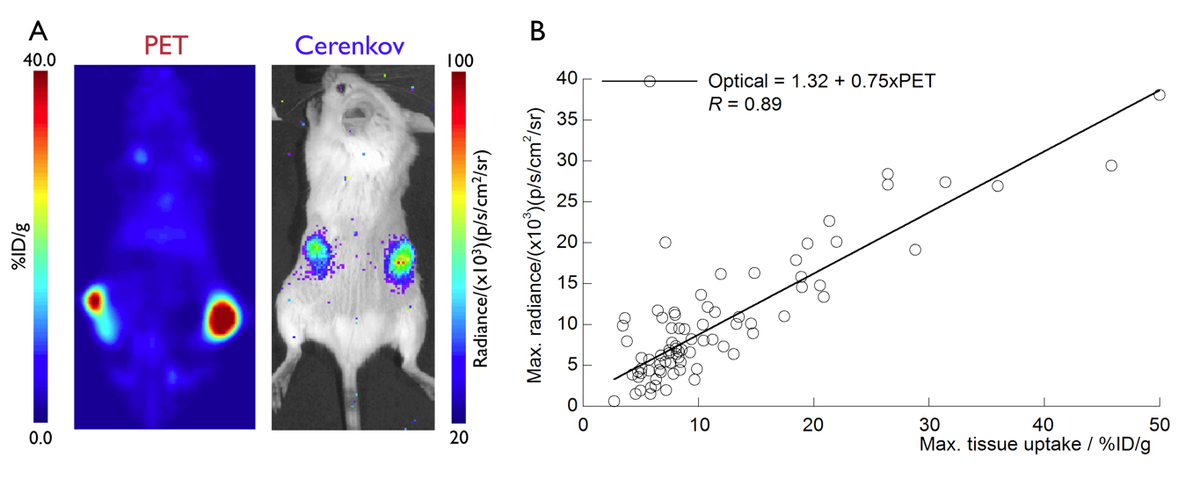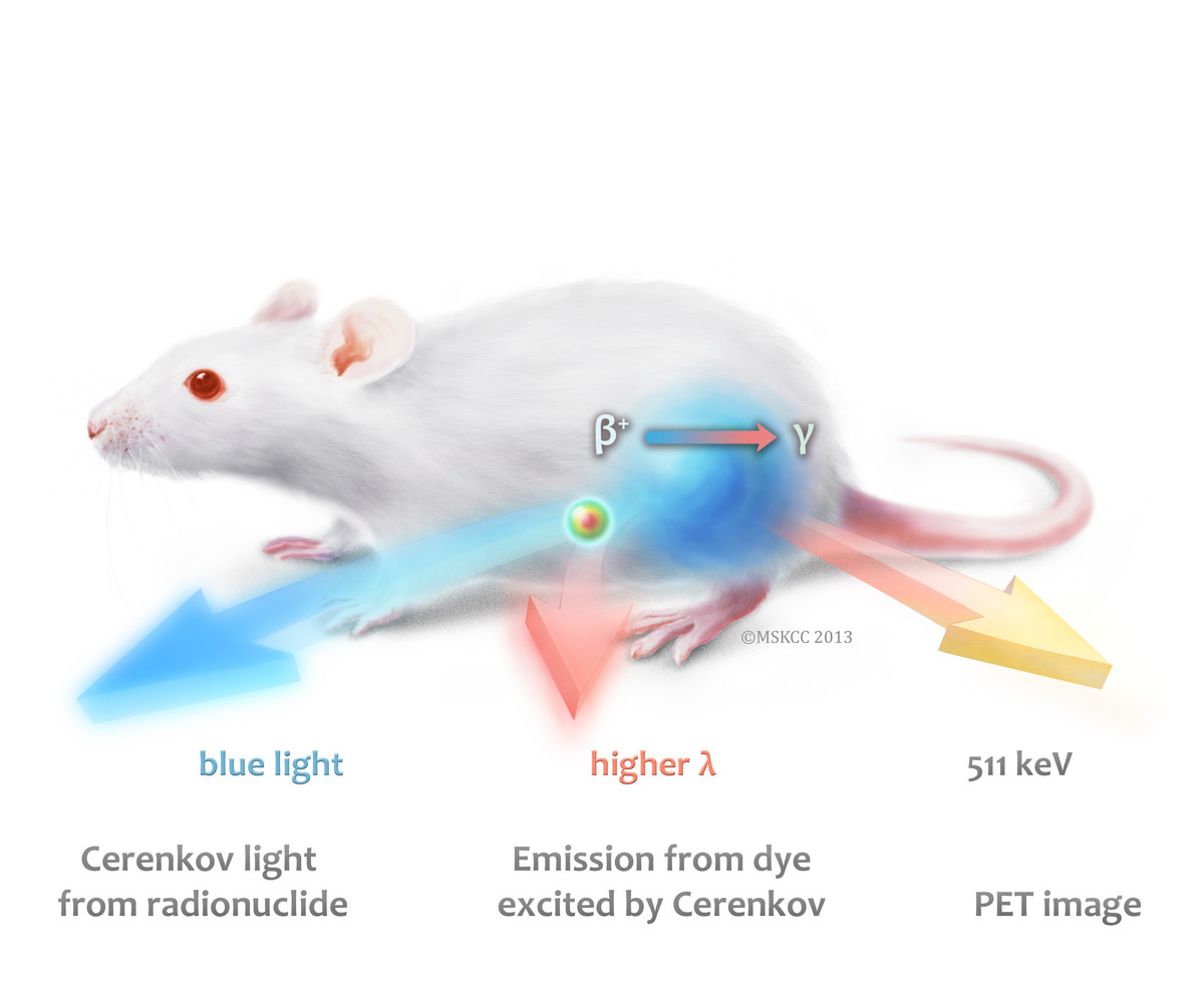
Cerenkov luminescence imaging (CLI) is an emerging hybrid modality that utilizes the light emission from many commonly used medical isotopes. Cerenkov radiation (CR) is produced when charged particles travel through a dielectric medium faster than the speed of light in that medium. First described in detail nearly 100 years ago, CR has only recently been applied for biomedical imaging purposes. The modality is of considerable interest as it enables the use of widespread luminescence imaging equipment to visualize clinical diagnostic (all PET radioisotopes) and many therapeutic radionuclides. The amount of light detected in CLI applications is significantly lower than in other optical imaging techniques such as bioluminescence and fluorescence. However, significant advantages in technology onthe camera detector makes CLI now an interesting methodology certainly for preclinical imaging and possibly also for clinical applications.
The advantages of CLI over other optical imaging agents include the use of approved radiotracers and lack of an incident light source, resulting in high signal to background ratios. As well, multiple subjects may be imaged concurrently (up to 5 in common bioluminescent equipment) and much faster than with PET.

We recently piublished on “Cerenkov 2.0”, the utilization of Cerenkov to interrogate different disease signatures using unique and novel imaging agents, which are excited by Cerenkov to indicate antigen expression or - as activatable imaging agents - even to demonstrate enzymatic activity. These agents are inherently quantitative and have a better signal-to noise ratio compared to conventional fluorescence imaging. They also allow for the simultaneous analysis of different molecular entities. This paper has been published online on September 8, 2013 in Nature Medicine.
Our lab is interested in various Cerenkov applications, from combining CLI with nanotechnology in the preclinical realm to clinical translation; currently we also have one active clinical trial exploring Cerenkov imaging in patients. From this trial we recently published the first results, demonstrating the feasibility of clinical Cerenkov imaging.


