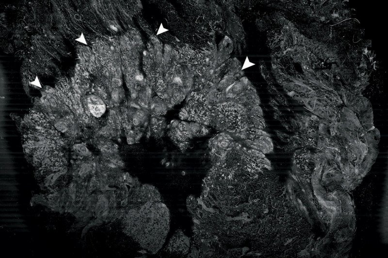
This image shows a cross section of a piece of tissue that was surgically removed from a patient with breast cancer and imaged using a method called confocal mosaicking microscopy. The arrowheads indicate the border between invasive cancer cells and the surrounding tissue.
A Memorial Sloan Kettering team of optical engineers and physicians is developing the technology to allow faster pathologic examination of tissue that could be used in the operating room to guide surgery. The method could enable surgeons to more accurately pinpoint tumors and completely remove them while making the procedure more effective, and could spare many patients from having repeat surgery, says optical engineer Milind Rajadhyaksha, who leads the project.
Waiting for Pathology
One potential application is breast-conserving surgery, in which a woman’s cancer is removed while sparing as much of the surrounding, normal breast as possible. To ensure that no tumor cells have been left behind, a margin of tissue around the tumor is usually sent from the operating room to a pathology lab to be processed and analyzed. Often the patient is sent home while the results are obtained, which usually takes several days.
If cancer cells are found in the margin tissue, she may need to return for more surgery. Repeat surgeries are not only burdensome for the patient but are associated with increased health risks and costs.
But Dr. Rajadhyaksha is hopeful that the microscope he and his colleagues are developing will eventually allow pathologists and surgeons to work in a more integrated way so that removal of the tumor and any additional tissue can be done in one session.
Mosaicking with a Laser Microscope
“The instrument operates by scanning a focused spot of laser light onto a piece of tissue and capturing the reflected and fluorescent light through a pinhole,” Dr. Rajadhyaksha explains. This produces crisp and detailed images of cells and tissue structures at different depths within the specimen. The images carry information equivalent to that obtained by traditional pathology.
“The main challenge is to make confocal imaging fast enough,” he adds. “To use the instrument in the operating room, clinicians need to be able to localize a tumor’s margins in few minutes.”
To increase the speed of imaging, the researchers came up with an approach called strip mosaicking, in which strips of the specimen are imaged individually and then “stitched” together by computer to create an image representing the entire cross section of the specimen. The analysis can now be done in 90 seconds on a square centimeter of surgically excised tissue.
“Skin surgical applications of this technology are already being explored in clinical trials in Europe,” says Dr. Rajadhyaksha, “and we hope to soon start testing it here at Sloan Kettering for surgery of the breast, head and neck areas, and non-melanoma skin cancers.”


