Molecular Biology Program
The Kenneth Marians Lab
Research

Kenneth J. Marians, PhD
William E. Snee Chair
Professor
Professor
Research in my laboratory centers on: (i) the events that occur when the replisome (the multi-component enzyme machine that replicates chromosomal DNA) encounters blockages to replication fork progression, and (ii) the mechanisms by which chromosomes are condensed and separated during cell division. Our studies on the consequences of replication fork blockage and restart utilize purified replication, recombination, and transcription proteins in various well-defined systems reconstituted in vitro that model all steps of these events. Our studies on chromosome condensation and segregation utilize a combination of biochemical, cell biological, and molecular genetic approaches.
Research Projects

Featured News
Feature
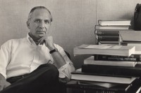
MSK colleagues pay tribute to molecular biologist Jerard Hurwitz’s scientific accomplishments and passion for discovery.
Q&A
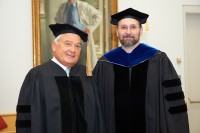
Meet the man who helped make the Gerstner Sloan Kettering Graduate School of Biomedical Sciences a reality.
In the News
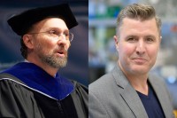
Cell biologist Michael Overholtzer will succeed DNA replication expert Ken Marians.
People
Kenneth J. Marians, PhD
William E. Snee Chair
Professor
- Kenneth Marians focuses on mechanisms of replication restart and chromosome segregation.
- PhD, Cornell University
- [email protected]
- Email Address
- 212-639-5890
- Office Phone
Members


Research Associate
Senior Research Assistant

Research Scholar
Research Scholar
Senior Research Assistant


Senior Research Scientist
Senior Research Technician
Postdoctoral Fellow
Postdoctoral Fellow
Research Fellow

Graduate Student
Graduate Student
Graduate Student
Research Fellow
