Memorial Sloan Kettering researchers have shed light on the process by which developing nerve cells, or neurons, are directed to split into two distinct parts — a long, slender axon that conducts electrical impulses away from a cell, and shorter dendrites that receive signals from other cells and conduct them into the cell body.
Investigators in the laboratory of developmental neurobiologist Songhai Shi gained an important insight into how a protein called mPar3 guides this asymmetrical formation of a neuron, known as neuronal polarization. Axon/dendrite polarity is essential for the one-directional flow of information in the nervous system.
The mPar3 protein was found to regulate microtubules — microscopic, threadlike structures inside cells that help them maintain shape and movement. Although it was already known that mPar3 plays a key role in neuronal polarization, the discovery that the protein directs this process through microtubule regulation was unexpected.
“This is the first time that mPar3 has been linked to microtubules and this relationship has been shown to be critical to proper development of a nerve cell,” says Dr. Shi, the senior author.
The study, reported in the January 14 issue of Developmental Cell, was led by She Chen, who recently completed a fellowship in Dr. Shi’s laboratory.
Unbalanced Activity
mPar3 is the mammalian form of the Par3 protein, which has long been recognized as an important factor in cell polarity from studies in a variety of animals, including worms, fruit flies, and mice. Earlier research performed in mice by Dr. Shi and others found that during embryonic development mPar3 accumulates in the part of the neuron destined to become an axon, suggesting that the protein spurs axon growth and neuron polarization. But it was unclear how mPar3 produces this outcome.
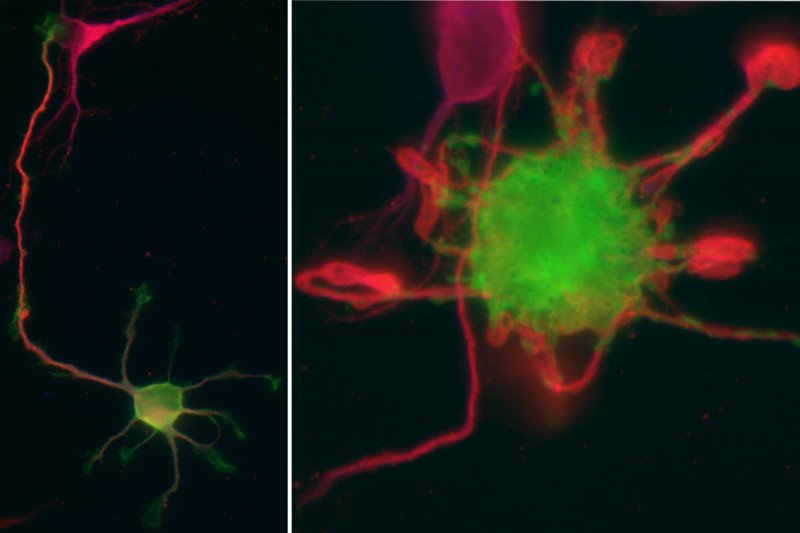
In the latest study, the researchers analyzed embryonic mouse brain cells in culture and found that mPar3 guides polarization by selectively binding to, bundling, and stabilizing microtubules in particular parts of the neuron at specific times. Increased microtubule stabilization at one site, such as the region that gives rise to the axon, allows that part of the cell to grow faster than other regions.
When mPar3’s regulation of microtubules is disrupted, the neuron cannot correctly differentiate into axon and dendrites. The researchers demonstrated this by blocking and boosting mPar3 function, which in both cases resulted in improperly developed neurons.
Polarization of Human Cells
Dr. Shi says that his lab’s discoveries could have broad implications beyond the development of neurons.
“Both mPar3 and microtubules have been conserved by evolution across many species, so this relationship is likely to hold true for polarization in other cell types, including human cells,” he explains.
